Dental X-ray positioning is a critical technique in modern dentistry‚ enabling accurate diagnoses and effective treatment planning. Proper positioning ensures clear‚ detailed images of dental structures‚ guiding precise care.
1.1 Importance of Proper X-Ray Positioning in Dentistry
Proper X-ray positioning is essential for accurate diagnoses and effective treatment planning in dentistry. It ensures clear‚ detailed images of dental structures‚ minimizing retakes and radiation exposure. Correct alignment of the X-ray beam with the film or sensor guarantees diagnostic accuracy‚ reducing errors in identifying conditions like dental caries or periodontal disease. Proper positioning also enhances patient safety by optimizing radiation dose and image quality. Specialized tools‚ such as positioning kits‚ help achieve precise alignment‚ ensuring consistency and reliability in radiographic outcomes. This fundamental step is critical for delivering high-quality dental care and maintaining patient trust in diagnostic procedures.
1.2 Overview of Dental X-Ray Types
Dental X-rays are categorized into intraoral and extraoral types‚ each serving specific diagnostic purposes. Intraoral X-rays‚ such as periapical and bitewing radiographs‚ capture detailed images of individual teeth and surrounding structures. Extraoral X-rays‚ including panoramic and cephalometric radiographs‚ provide broader views of the jaw and facial bones. Panoramic X-rays offer a two-dimensional image of the entire mouth‚ while cephalometric X-rays are used for orthodontic assessments. Cone-beam CT (CBCT) is an advanced type‚ offering three-dimensional imaging for complex cases. Each type requires precise positioning to ensure accurate results‚ making them invaluable tools in modern dental diagnostics and treatment planning.
Radiation Safety in Dental X-Ray Positioning
Radiation safety in dental X-ray positioning focuses on minimizing exposure while ensuring diagnostic image quality‚ protecting both patients and dental staff from unnecessary radiation.
2.1 Principles of Radiation Protection
The principles of radiation protection in dental X-ray positioning emphasize minimizing exposure through justification‚ optimization‚ and dose limitation; Justification ensures that radiographs are taken only when necessary. Optimization involves using the lowest possible dose to achieve diagnostic-quality images. Dose limitation restricts exposure to levels considered safe for patients and staff. Proper shielding‚ such as lead aprons and thyroid collars‚ is essential. Additionally‚ digital radiography systems‚ which reduce radiation doses compared to traditional film‚ are widely recommended. Regular monitoring and training for dental professionals further enhance safety protocols‚ ensuring compliance with regulatory guidelines and patient protection.
2.2 Patient Selection for Radiographic Examinations
Patient selection for radiographic examinations is based on clinical judgment and the assessment of individual needs. Factors such as symptoms‚ risk of caries‚ periodontal disease‚ and dental implant placement guide the decision. Adults with good oral health and low risk may require X-rays every 24-36 months‚ while high-risk patients may need more frequent imaging. Children and adolescents‚ due to developing dentition‚ often require tailored approaches. Patient history‚ including previous dental work and systemic conditions‚ is considered. Modern technologies like digital radiography reduce radiation exposure‚ making imaging safer and more accessible. Proper patient selection ensures that radiographs are taken only when necessary‚ adhering to principles of radiation protection and diagnostic yield.
2.3 Safety Guidelines for Dental Radiography
Ensuring radiation safety is paramount in dental radiography. Guidelines emphasize minimizing exposure through proper techniques and equipment. Digital sensors and film placement reduce radiation doses. Lead aprons and thyroid collars are essential for patient protection. The ALARA principle (As Low As Reasonably Achievable) guides exposure levels. Regular maintenance of X-ray machines and sensors ensures optimal performance. Training staff in radiation safety protocols is critical. Patients should be educated on the benefits and risks of radiography. Strict adherence to safety measures minimizes health risks while maximizing diagnostic value. Compliance with regulatory standards ensures a safe environment for both patients and practitioners.

Dental X-Ray Positioning Guide
A comprehensive guide to dental X-ray positioning ensures accurate imaging and patient safety. It covers intraoral and extraoral techniques‚ patient preparation‚ and proper equipment alignment for optimal results.
3.1 Intraoral X-Ray Positioning Techniques
Intraoral X-ray positioning involves placing the film or digital sensor inside the patient’s mouth. Proper alignment of the X-ray beam with the dental structures is essential for clear images. Techniques like the paralleling method ensure the beam is perpendicular to the film‚ reducing distortion. The bisecting angle technique angles the beam to bisect the angle between the dental structure and the film. Both methods aim to provide accurate representations of teeth and surrounding tissues. Proper patient preparation‚ such as positioning the head and ensuring the sensor is securely placed‚ minimizes movement and ensures diagnostic-quality images while adhering to radiation safety protocols.
3.2 Extraoral X-Ray Positioning Techniques
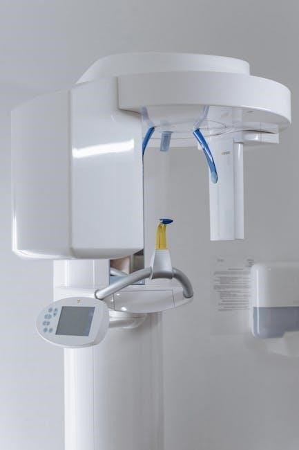
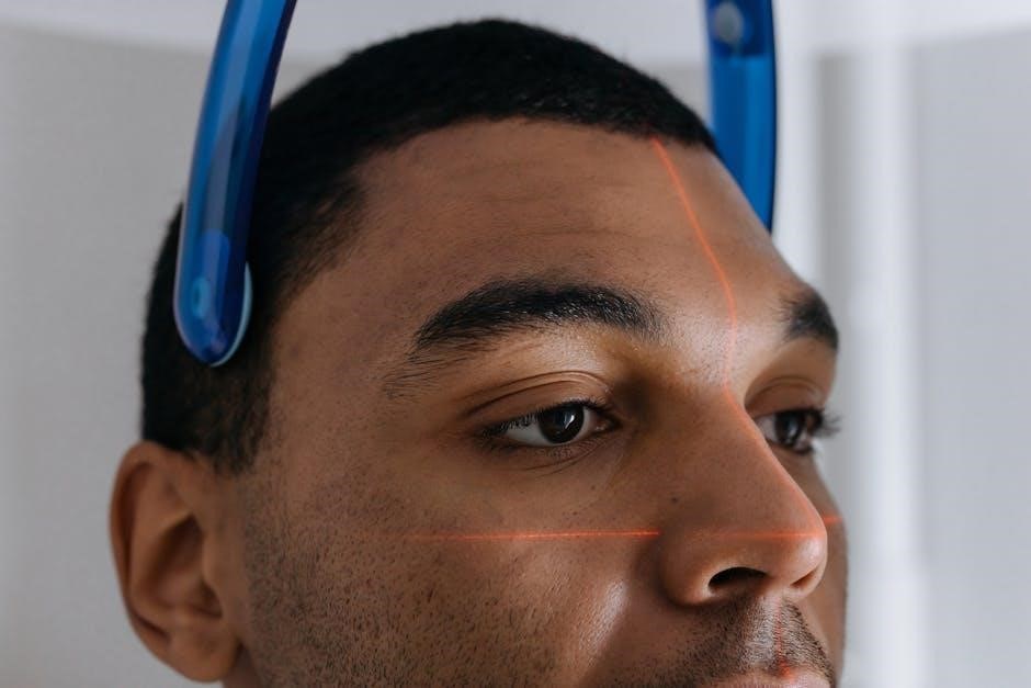
Extraoral X-ray positioning involves capturing images with the film or sensor outside the mouth. Techniques like panoramic radiography require the patient to stand upright with the head positioned so the X-ray beam rotates around the skull. Cephalometric radiography involves side-view imaging for profiling facial structures. Proper alignment ensures the beam passes through the midline of the face. CBCT positioning may involve specific head rests and chin supports to maintain stability. Each technique requires precise patient positioning to achieve accurate‚ distortion-free images. Proper alignment and immobilization are crucial for diagnostic clarity while minimizing radiation exposure‚ ensuring safe and effective imaging for comprehensive dental assessments.
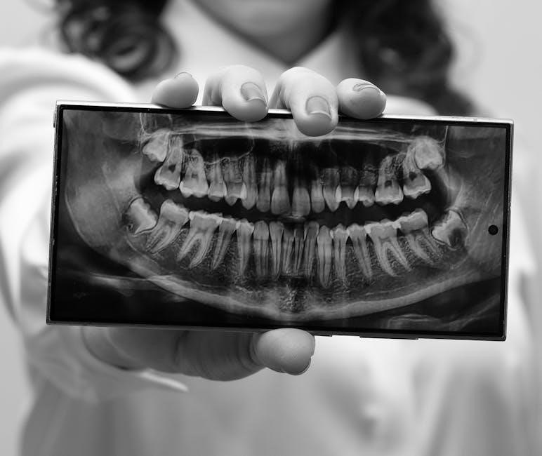
3.3 Panoramic X-Ray Positioning
Panoramic X-ray positioning captures a wide view of the dental and facial structures in a single image. The patient stands upright with the head positioned so the X-ray beam rotates around the skull. The chin is placed on a rest‚ and the head is aligned with the midline. The X-ray beam moves in a semicircular path to image the entire mouth‚ including teeth‚ jaws‚ and surrounding tissues. Proper positioning ensures the beam is perpendicular to the film‚ minimizing distortion. This technique is ideal for assessing the TMJ‚ sinus areas‚ and overall dental anatomy. It offers a comprehensive view with reduced radiation exposure compared to intraoral methods.
Intraoral X-Ray Positioning
Intraoral X-ray positioning involves placing the sensor inside the mouth to capture detailed images of individual teeth and surrounding structures. Proper angulation ensures clear‚ diagnostic-quality results.
4.1 Periapical Radiography Positioning
Periapical radiography focuses on capturing detailed images of individual teeth and their surrounding bone structure. The X-ray beam is directed at a specific angle to ensure the entire tooth‚ from crown to root‚ is visible. Proper positioning involves aligning the beam perpendicular to the dental film or sensor‚ placed inside the patient’s mouth. This technique minimizes distortion and provides a clear view of the tooth’s anatomy‚ aiding in diagnosing conditions like decay‚ abscesses‚ or bone loss. Accurate positioning is critical for obtaining diagnostic-quality images‚ ensuring precise treatment planning and patient care.
4.2 Bitewing Radiography Positioning
Bitewing radiography involves positioning the X-ray beam to capture images of the upper and lower teeth biting down on a radiographic film or sensor. This technique is essential for detecting interproximal caries‚ assessing the marginal fit of restorations‚ and monitoring periodontal changes. Proper positioning requires the beam to be angled at 10-15 degrees from the vertical‚ ensuring that the film captures both the upper and lower teeth in a single image. The patient is asked to bite gently on the film to hold it in place‚ ensuring clarity and accuracy. This method is crucial for early detection of dental issues and effective treatment planning.
4.3 Occlusal Radiography Positioning
Occlusal radiography involves positioning the X-ray beam to capture detailed images of the maxillary or mandibular arch. The technique requires placing the film or sensor on the occlusal surface of the teeth‚ with the X-ray beam directed perpendicular to the film. This positioning is ideal for assessing the floor of the mouth‚ locating retained teeth‚ or evaluating large lesions. Proper alignment ensures minimal distortion and clear visualization of anatomical structures. The patient is instructed to close their mouth gently to hold the film in place‚ ensuring optimal image quality. Occlusal radiographs are particularly useful for diagnosing conditions affecting the broader dental arch and surrounding tissues.
Extraoral X-Ray Positioning
Extraoral X-ray positioning involves placing the X-ray source outside the mouth to capture images of larger dental structures. It is used for comprehensive assessments of the maxillofacial region‚ providing a broader view of the teeth‚ jaws‚ and surrounding tissues. This technique is particularly useful for evaluating the temporomandibular joint (TMJ)‚ jaw fractures‚ and other complex conditions.
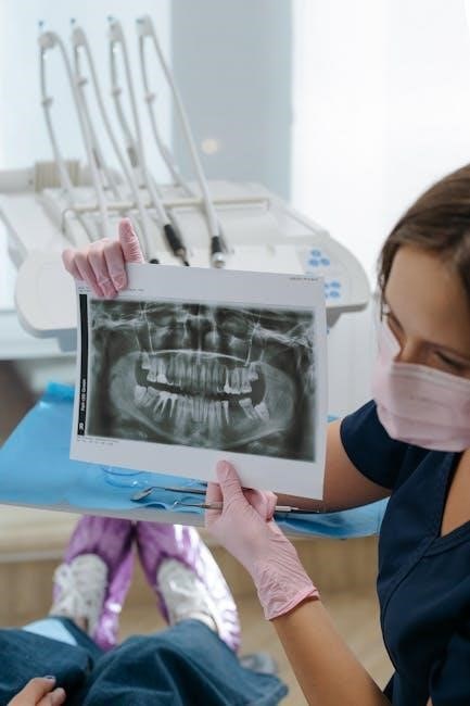
5.1 Panoramic Radiography Positioning
Panoramic radiography positioning involves capturing a two-dimensional image of the entire mouth in a single X-ray. The patient stands upright or sits comfortably‚ with the tongue pressed against the palate and lips relaxed. A bite block is often used to stabilize the jaw. The X-ray beam rotates around the head‚ creating a wide view of the teeth‚ upper and lower jaws‚ and surrounding facial bones. Proper alignment ensures minimal distortion and clear visualization of the maxillofacial region. This technique is ideal for assessing the overall dental structure‚ temporomandibular joint (TMJ)‚ and jaw fractures‚ providing a comprehensive diagnostic tool for complex cases.
5.2 Cephalometric Radiography Positioning
Cephalometric radiography positioning involves capturing lateral or frontal images of the skull and facial bones. The patient stands or sits upright with the head in a natural‚ neutral position. The Frankfort horizontal plane is often used as a reference‚ aligning the lower margin of the left orbit and the upper margin of the left external auditory meatus. The midsagittal plane is centered perpendicular to the X-ray beam. The beam is directed horizontally‚ ensuring a parallel relationship to the floor and perpendicular to the midsagittal plane. Proper positioning minimizes distortion‚ allowing accurate assessment of craniofacial structures for orthodontic and surgical planning. This technique is vital for evaluating growth patterns and treatment progress in orthodontics.

Equipment for Dental X-Ray Positioning
Dental X-ray positioning requires specialized equipment‚ including X-ray machines‚ digital sensors‚ and film holders. These tools ensure accurate imaging and proper alignment for diagnostic clarity and safety.
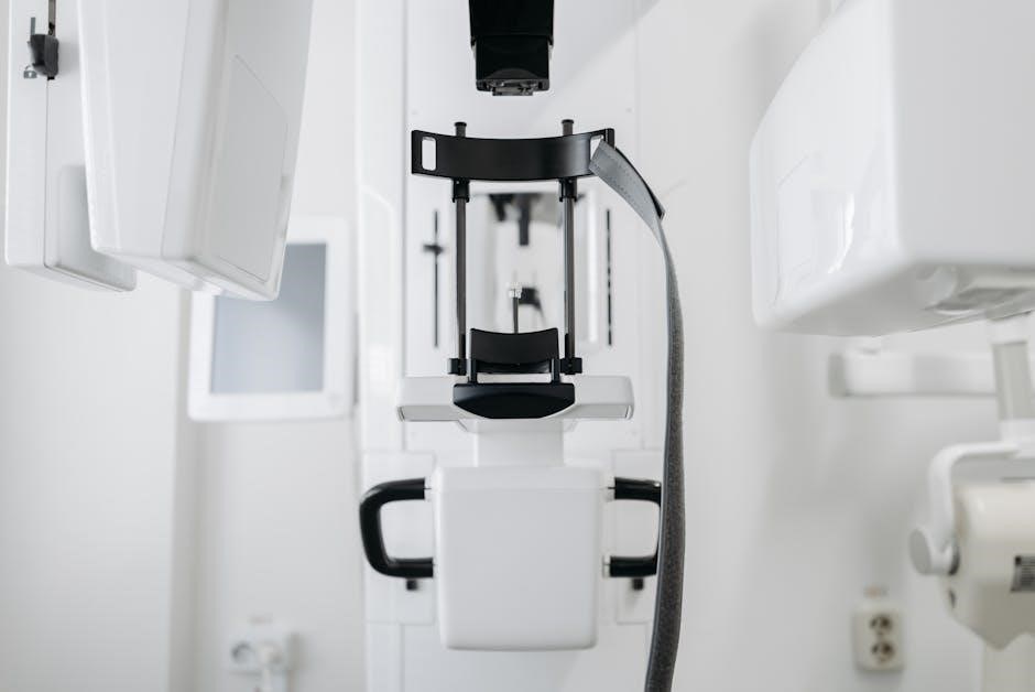
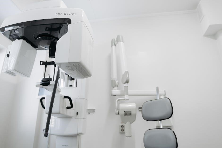
6.1 X-Ray Machine and Accessories
The X-ray machine is the core of dental radiography‚ producing controlled radiation for imaging. Accessories like digital sensors‚ film holders‚ and positioning guides enhance accuracy and patient comfort. These tools ensure proper alignment of the X-ray beam with the target area‚ minimizing retakes and radiation exposure. Advanced machines often feature adjustable settings to optimize image quality based on patient needs. Accessories such as radiopaque markers help in accurate positioning‚ while film or sensor holders maintain stability during exposure. Together‚ the machine and its accessories are essential for capturing clear‚ diagnostic images that aid in precise treatment planning and patient care.
6.2 Digital Sensors and Film Placement
Digital sensors and film placement are critical for obtaining high-quality radiographic images. Proper positioning ensures accurate representation of dental structures‚ minimizing distortion. Digital sensors offer instant results and reduced radiation exposure compared to traditional film. Film placement requires precise alignment with the X-ray beam to capture detailed anatomy; Incorrect positioning can lead to retakes‚ increasing radiation exposure and diagnostic delays. Accessory tools‚ such as bite blocks and positioning guides‚ help stabilize the sensor or film‚ ensuring optimal alignment. Proper placement is essential for diagnostic accuracy‚ patient safety‚ and efficient workflow in dental radiography.
Patient Preparation for X-Ray Positioning
Patient preparation is vital for effective dental X-ray positioning. It ensures comfort‚ safety‚ and accurate imaging. Proper alignment and positioning minimize radiation exposure and improve diagnostic outcomes.
7.1 Patient Positioning in the Dental Chair
Patient positioning in the dental chair is crucial for accurate X-ray imaging. The patient should be seated comfortably‚ with their head aligned to ensure proper angulation of the X-ray beam. For intraoral X-rays‚ the patient’s mouth is opened slightly‚ and the sensor or film is placed correctly. The dentist adjusts the chair to position the patient’s head at the correct height and angle‚ ensuring the X-ray beam is perpendicular to the dental film or sensor. Proper alignment minimizes distortion and ensures clear‚ diagnostic-quality images. Clear communication and guidance help the patient remain still during the procedure‚ reducing movement and improving outcomes. This step is essential for obtaining precise radiographic results.
7.2 Aligning the X-Ray Beam
Aligning the X-ray beam correctly is essential for obtaining accurate and diagnostic-quality images. The central X-ray beam should be directed perpendicular to the dental film or sensor‚ ensuring proper coverage of the desired anatomical structures. For intraoral X-rays‚ the beam is typically aimed at the apical region of the teeth‚ while for extraoral X-rays‚ the beam is adjusted to capture the broader facial structures. Proper alignment minimizes distortion and ensures that the resulting image is clear and detailed. Adjustments may be made based on the specific procedure and patient anatomy to optimize image quality and diagnostic value. This step is critical for precise radiographic outcomes.
Quality Assurance in Dental Radiography
Quality assurance ensures consistent image quality‚ minimizing retakes and errors. It involves regular equipment maintenance‚ proper techniques‚ and adherence to radiation safety protocols.
8.1 Ensuring Image Quality
Ensuring image quality in dental radiography is crucial for accurate diagnoses and effective treatment planning. Proper positioning techniques‚ such as using the iM3 X-Ray Positioning Kit‚ help achieve clear and detailed images. Correct alignment of the X-ray beam‚ ensuring it passes through the target area‚ and precise placement of digital sensors are essential. Adhering to established guidelines and maintaining equipment properly also contribute to consistent image quality. Regular quality assurance checks‚ proper training for dental staff‚ and continuous monitoring of imaging processes further enhance the reliability of radiographic images‚ ensuring optimal patient care and successful treatment outcomes.
8.2 Reducing Retakes and Errors
Reducing retakes and errors in dental radiography is essential for improving efficiency and patient care. Proper training and adherence to established positioning guidelines are critical. Ensuring the X-ray beam is correctly aligned with the digital sensor or film‚ and the patient is positioned accurately‚ minimizes the need for retakes. Regular equipment maintenance and quality assurance programs also play a key role. Additionally‚ using tools like the iM3 X-Ray Positioning Kit can help standardize techniques. By following these steps‚ dental professionals can reduce errors‚ enhance image quality‚ and ensure patient safety while maintaining compliance with radiation safety standards and regulatory requirements. This approach streamlines workflows and improves overall outcomes.

Clinical Applications of Proper X-Ray Positioning
Proper X-ray positioning aids in diagnosing dental caries‚ assessing periodontal disease‚ and evaluating dental implants‚ ensuring accurate and effective treatment planning for various dental conditions.
9.1 Diagnosing Dental Caries
Dental caries‚ often detected through X-rays‚ involve tooth decay visible as darkened areas. Proper positioning ensures clear images‚ helping identify early stages of decay for timely intervention‚ preventing progression and preserving tooth structure. Accurate X-ray placement is crucial for diagnosing caries‚ especially in areas invisible to the naked eye‚ such as between teeth or beneath restorations. By capturing detailed images‚ dentists can assess the extent of decay and plan appropriate treatments‚ whether fillings‚ crowns‚ or more complex procedures. Early detection through precise X-ray positioning significantly improves patient outcomes and reduces the need for extensive dental work.
9.2 Assessing Periodontal Disease
Periodontal disease‚ affecting the gums and bone supporting teeth‚ is often assessed via X-rays to evaluate bone loss and tissue changes. Proper positioning ensures detailed images of the alveolar bone‚ enabling accurate measurement of pocket depths and detecting early signs of disease. X-rays reveal the extent of bone resorption‚ helping diagnose the severity of periodontitis. This information is crucial for developing treatment plans‚ such as scaling‚ root planing‚ or surgical interventions. Regular radiographic assessments also monitor disease progression and response to therapy‚ ensuring effective long-term management of periodontal health. Accurate X-ray positioning is essential for reliable diagnostic outcomes in periodontal care.
9.3 Evaluating Dental Implants
Dental implants require precise radiographic evaluation to assess their placement‚ integration‚ and long-term success. X-rays provide critical insights into the relationship between the implant and surrounding bone‚ ensuring proper osseointegration. Proper positioning techniques are essential to obtain clear images of the implant’s length‚ angle‚ and proximity to adjacent structures. Radiographs help identify potential complications‚ such as bone resorption or implant malposition‚ allowing for timely interventions. Regular X-ray evaluations also monitor the healing process and stability of the implant over time. Accurate imaging is vital for ensuring the success and longevity of dental implants‚ making X-ray positioning a cornerstone of implant assessment and management.
Legal and Regulatory Considerations
Compliance with radiation safety regulations is essential for dental practices. Proper documentation and record-keeping ensure adherence to legal standards‚ safeguarding patient welfare and professional accountability.
10.1 Compliance with Radiation Safety Regulations
Compliance with radiation safety regulations is paramount in dental practices to ensure patient and staff protection. Regulations mandate the use of proper equipment‚ techniques‚ and dosages to minimize exposure. Dental professionals must adhere to guidelines set by governing bodies‚ such as the Department of Health‚ to maintain safety standards. Regular training and updates on radiation protocols are essential for staff. Proper documentation and record-keeping are also required to demonstrate adherence to legal standards. Non-compliance can result in legal consequences‚ emphasizing the importance of strict radiation safety practices in all dental radiographic procedures.
10.2 Record-Keeping and Documentation
Accurate and thorough record-keeping is essential in dental radiography for legal‚ safety‚ and patient care purposes. Documentation includes details of each radiographic examination‚ such as the type of X-ray‚ patient information‚ and radiation exposure levels. Maintaining these records ensures compliance with regulatory requirements and provides a reference for future treatments. Digital systems are increasingly used for efficient storage and retrieval of radiographic data. Proper documentation also aids in tracking patient history and supports continuity of care. Additionally‚ it serves as a protective measure for dental practices‚ offering evidence of adherence to safety protocols and professional standards.
Advanced Techniques in Dental X-Ray Positioning
Advanced techniques like Cone-Beam CT (CBCT) and 3D imaging revolutionize dental radiography‚ offering detailed visuals for precise diagnostics and treatment planning‚ enhancing traditional X-ray methods significantly;
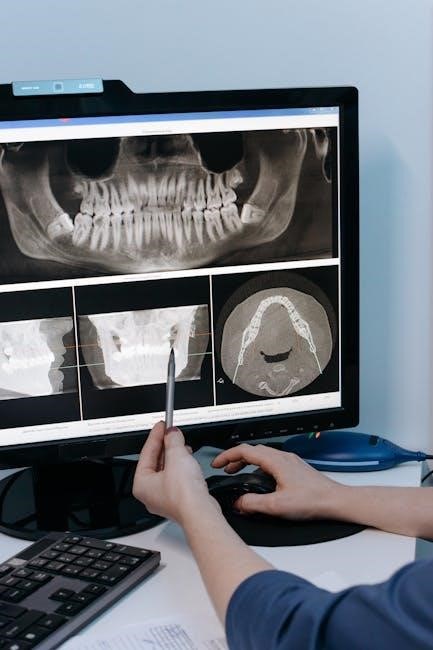
11.1 Cone-Beam CT (CBCT) Positioning
Cone-Beam CT (CBCT) positioning is an advanced imaging technique providing three-dimensional visualization of dental structures. It is particularly useful for evaluating complex anatomical details‚ such as dental implants‚ bone density‚ and periodontal disease. Proper patient positioning involves aligning the region of interest with the CBCT machine’s focal point to ensure accurate and detailed images. The technique minimizes distortion and offers superior diagnostic capabilities compared to traditional 2D radiography. CBCT is also valuable for orthodontic assessments and surgical planning‚ ensuring precise treatment outcomes. Proper positioning is critical to achieve high-quality images and optimize diagnostic accuracy in modern dental practices.
11.2 3D Imaging in Dental Radiography
3D imaging in dental radiography revolutionizes diagnostic capabilities by providing detailed‚ three-dimensional views of oral structures. Unlike traditional 2D imaging‚ 3D techniques‚ such as cone-beam CT‚ capture complex anatomy with precision. This enhances the ability to diagnose conditions like fractures‚ cysts‚ and bone defects. 3D imaging also aids in treatment planning for dental implants‚ orthodontic cases‚ and surgical procedures. Proper positioning is essential to ensure accurate and comprehensive imaging. The technology reduces errors in diagnosis and improves patient outcomes. By offering a holistic view of dental and facial structures‚ 3D imaging has become a cornerstone of modern‚ advanced dental radiography‚ elevating both diagnostic accuracy and treatment efficiency.
12.1 Summary of Key Positioning Principles
Proper dental X-ray positioning ensures accurate imaging‚ minimizing distortion and radiation exposure. Key principles include aligning the X-ray beam perpendicular to the dental film or sensor‚ using techniques like paralleling or bisecting for intraoral views‚ and maintaining the correct vertical and horizontal angles. Patient preparation‚ such as proper seating and positioning in the dental chair‚ is crucial for optimal results. Digital sensors and film placement must be precise to capture detailed images. Adhering to radiation safety guidelines‚ such as using lead aprons and thyroid collars‚ balances image quality with patient protection. These principles ensure diagnostic accuracy‚ supporting effective treatment planning and patient care.
12.2 Future Trends in Dental X-Ray Technology
Advancements in dental X-ray technology are revolutionizing imaging practices. Cone-beam CT (CBCT) and 3D imaging are becoming more prevalent‚ offering detailed anatomical views for complex diagnoses. Artificial intelligence (AI) is being integrated to enhance image analysis‚ improve diagnostic accuracy‚ and automate positioning techniques. Future trends also include the development of lower-dose X-ray systems‚ reducing radiation exposure while maintaining image quality. Additionally‚ the integration of digital sensors with advanced software promises real-time imaging and faster processing. These innovations aim to improve patient outcomes‚ streamline workflows‚ and set new standards for dental radiography‚ ensuring safer‚ more efficient‚ and precise imaging solutions.